University of Massachusetts becomes the first hospital in the United States to use the Atlas Stent for Stent-Assisted Coiling of Cerebral Aneurysm
Date Posted: Saturday, August 01, 2015On June 30, 2015, Dr. Ajit S. Puri became the first physician in the United States to use the Atlas Stent for cerebral aneurysm stent-assisted coiling treatment for one of our neurointerventional patients. Endovascular treatment of cerebral aneurysms is a minimally invasive technique that has evolved rapidly since the first detachable coils were used to treat a patient in 1991. The procedure involves accessing the brain’s blood vessels with tiny plastic catheters introduced through a small puncture in the groin. The embolization coils used to treat the aneurysm are made of a metal that has a memory so that when the coils are advanced into the aneurysm, the metal unfolds, filling the space within the aneurysm. Advanced techniques in endovascular therapy have evolved to allow for the use of stents, similar in concept to the types of devices used to treat coronary artery disease, as an adjunctive therapy to treat wide-neck aneurysms. The stents serve as scaffolding for coil placement and prevent intrusion into the “parent” artery while allowing for the maximum number of coils to be deployed within the aneurysm.
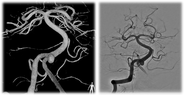 Figure 1-Pretreatment images of the aneurysm |
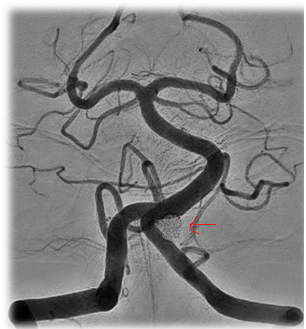 Figure 3: Final angiographic result. Note the red arrows showing the final coil mass. |
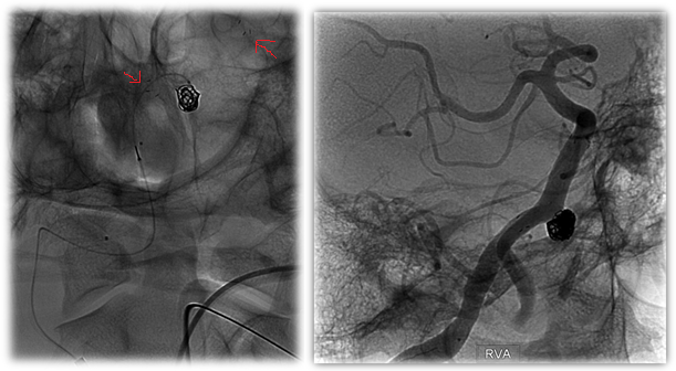 Figure 2: Intraprocedural Images of First Atlas Case. Note the red arrows indicating the radiopaque markers on either end of the stent. |
These very small stents are made of nitinol metal alloy that are cut by a laser in a specific pattern to optimize the contact with the blood vessel wall. Generally speaking, there are two design patterns to describe these types of devices, open-cell and closed-cell, and this refers to the configuration of the metal struts within the stent.
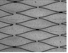 Figure 4: Open-Cell Stent Design |
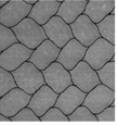 Figure 5: Closed-cell stent Design |
Recently, Stryker Neurovascular developed a refined version of this type of stent called the Atlas Neuroform Stent System. This version of the stent uses a hybrid design that combines both types of cell arrangements to maximize the navigability and conformability of the stent while preserving the radial force needed to retain the coils. This device is special because for the first time it can be delivered through the same small microcatheter that is used to place the coils. Previously, larger catheters were required. Now, it is feasible to place the stent, and then use the same microcatheter to subsequently coil the aneurysm behind the stent. This advancement is non-trivial from a technology perspective, and has the potential to benefit patients with small, atraumatic access in a simpler, faster procedure.
UMASS Radiology New England Center for Stroke Research, under the leadership of Drs. Gounis, Puri and Wakhloo, led the preclinical assessment and ultimate design validation of this new device over the past 2 years. It is fitting that Dr. Puri, with already years of experience with this new technology in animal models, was the first in the US to implant the device in a patient.


