Terarecon
Date Posted: Monday, January 30, 2017
This is an exciting time to participate in the future of our radiology department. The installation of TeraRecon is underway.
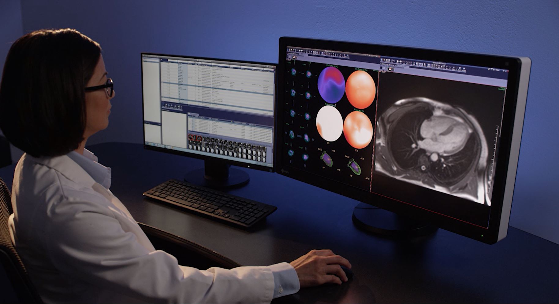
TeraRecon is a single solution tool that we hope will unify and simplify the myriad of software we are currently using for post processing of images. TeraRecon offers the ability to connect with all of our imaging modalities to improve productivity by utilizing one universal system. The current complex system of learning different tools for each of our scanners and post processing workstations results in fragmentation of skills and inefficiency for our technologists and radiologists. I am hopeful that the integration of these tools in one software package will enhance the clinical experience.
Advanced post processing tools including 3D clinical tools provided by TeraRecon holds the potential to improve diagnostic accuracy and the understanding of anatomy and pathology. This is in keeping with our missions to stand at the forefront of advanced imaging and teach our fellows, residents, medical students, and patients. TeraRecon offers the opportunity to simultaneously pull from many different Picture Archive and Communications systems (PACs) and Vendor Neutral Archives (VNA) in one organized timeline. As the department continues to grow, having a system that can pull from multiple different sources may significantly improve our diagnostic ability, providing real-time comparisons from different archives. This is an avenue to explore with our affiliations including Shields Radiology and Carewell. The ability to run the program directly off the server allows for flexibility and portability of the system with little demand or requirements on the individual desktop or laptop. This allows the radiologist to work from virtually anywhere with internet access.
One of the most interesting tools to explore is the advanced automation capability with preprocessing of multi-step 3D presentation states. For example, this program has the potential to preprocess a CTA of the neck with bone subtraction, curved planar reformats of the major cervical arterial vasculature, and NASCET calculation of ICA stenosis, prior to the radiologist or technologist opening the exam. This program also has ability to preprocess perfusion studies of the brain which should significantly improve turnaround time of perfusion stroke imaging.
I am looking forward to the opportunity fully explore the potentials of this program and would encourage those interested to join.
Andrew Chen
Assistant Professor of Neuroradiology


