iCels Innovations Lab – 3D Printing Cluster
Date Posted: Friday, March 18, 2016|
|
|
|
|
|
|
|
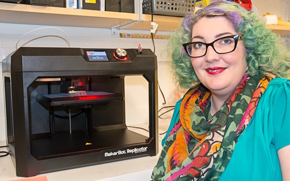
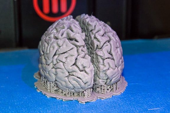
The first 3D prints are coming off the line in the newly established 3D Printing Cluster at the iCels Innovations Lab. Headed by Yasmin Carter Ph.D., recent faculty hire in the Division of Translational Anatomy, the 3D Printing Cluster aims to utilize new techniques and technologies to improve communication in education, patient interactions and research. Initial prints have been anatomical in nature and are created from CT and MRI images, objects can also be designed in 3D software programs.
Models for Medical Students
Dr. Carter plans to use models for teaching in the DSF course for the university where the first year medical students learn anatomy. One of her projects this summer is working with a student to create an anatomically functional and moveable model to study for sports medicine.
The 3D printers allow for personalization and detail not available with two-dimensional education materials. It also is reproducible, if a model is damaged or needs modifications it can be printed again at relatively low cost.
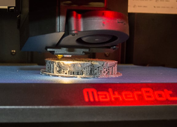
Currently, Dr. Carter is running two MakerBot 3D Printers. The first is a single extruder model and is able to print models faster but with less detail. The other model has two extruders which allows the use of two colors or two types of printing material, including dissolvable. Two colors may help to highlight different parts of anatomy or objects. The models can be painted after printing if desired.
Potential for Patient Education
Additionally, models can be a useful tool for patient education. In a current cluster project, Dr. Carter is working with Radiologists specializing in Mammography to develop models of different types of lumps that are detected during mammography. Whether these models help patients comprehension when discussing biopsy options will be tested.
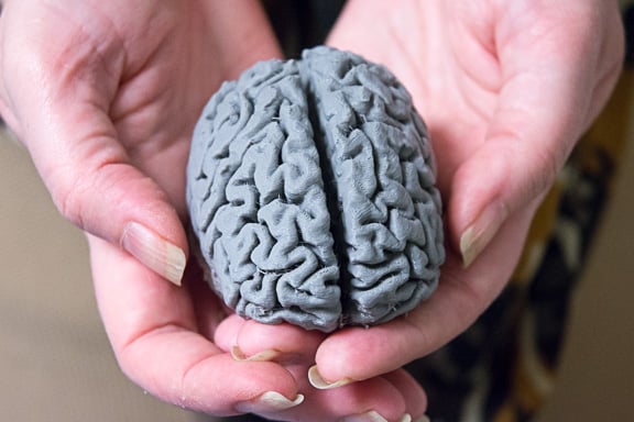
The models are built with several programs and then uploaded to the printer where it can take a few hours or days to print, depending on the size and complexity of the object. The quarter size brain pictured here took six hours to print.
Dr. Carter looks forward to future collaborations with both clinical and research faculty and invites interested parties to contact her.


