Eye & Retina
The Eye
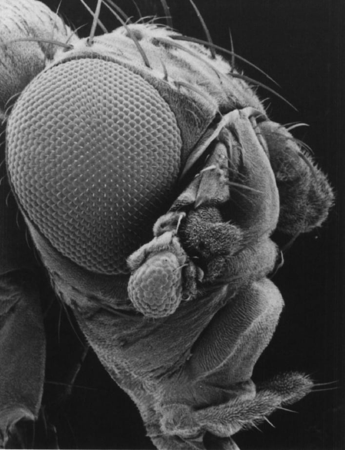
 The eye has always been an organ of controversy for evolutionary biologist. The difference in eye structure between a spider and a human is so stark that it seems illogical that both eyes should be evolutionarily related. It was therefore assumed, given the vast variety of eye structures found in nature, that the eye must have arisen by random chance more than 40 individual times during evolution. Charles Darwin knew that with his theory of evolution he was able to explain that once an eye was created by random chance then selective pressure would give rise to the diversity seen within different species. But within that logic was a basic problem. Opponents of Darwin's theory rightly pointed out that it seems to be impossible that such a perfect organ like the eye could have evolved by random chance more than 40 times. The solution for Darwin was to postulate that a simple prototype eye evolved only once very early in evolution, which then through natural selection could have given rise to the diversity present in nature. Modern molecular biology has now provided supportive evidence for such an idea. Genes that dictate eye development seem to be conserved throughout the animal kingdom suggesting that the birth of the eye was a single event in evolution.
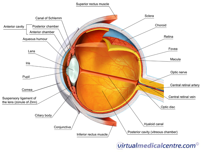
 Eyes allow animals to capture light and convert it into an electrical signal. However, while light is captured with our eyes the process of "seeing" an image occurs in our brain. The vertebrate eye is referred to as an "inverted camera" type eye as it is constructed much like a photo camera. Like in a camera the lens serves to focus light onto the retina, which is the photosensitive film in the back of our eyes, while the iris controls the amount of light that enters. The retina is a thin neuro-epithelial tissue that is comprised of 3 cellular layers. The name inverted however does not refer to the fact that the picture ends up being projected upside down onto the retina but rather refers to the arrangements of the different neurons within the retina. The photosensitive neurons of the retina that capture light are the furthest away from the incoming light source and thus point away from the light. Therefore, light has to cross first the entire retina before it can be captured by photoreceptors, the photosensitive neurons that serve to initiate the process of vision. Examples of non-inverted (meaning "verted") camera type eyes are the eye of the Cephalopods.
The Retina
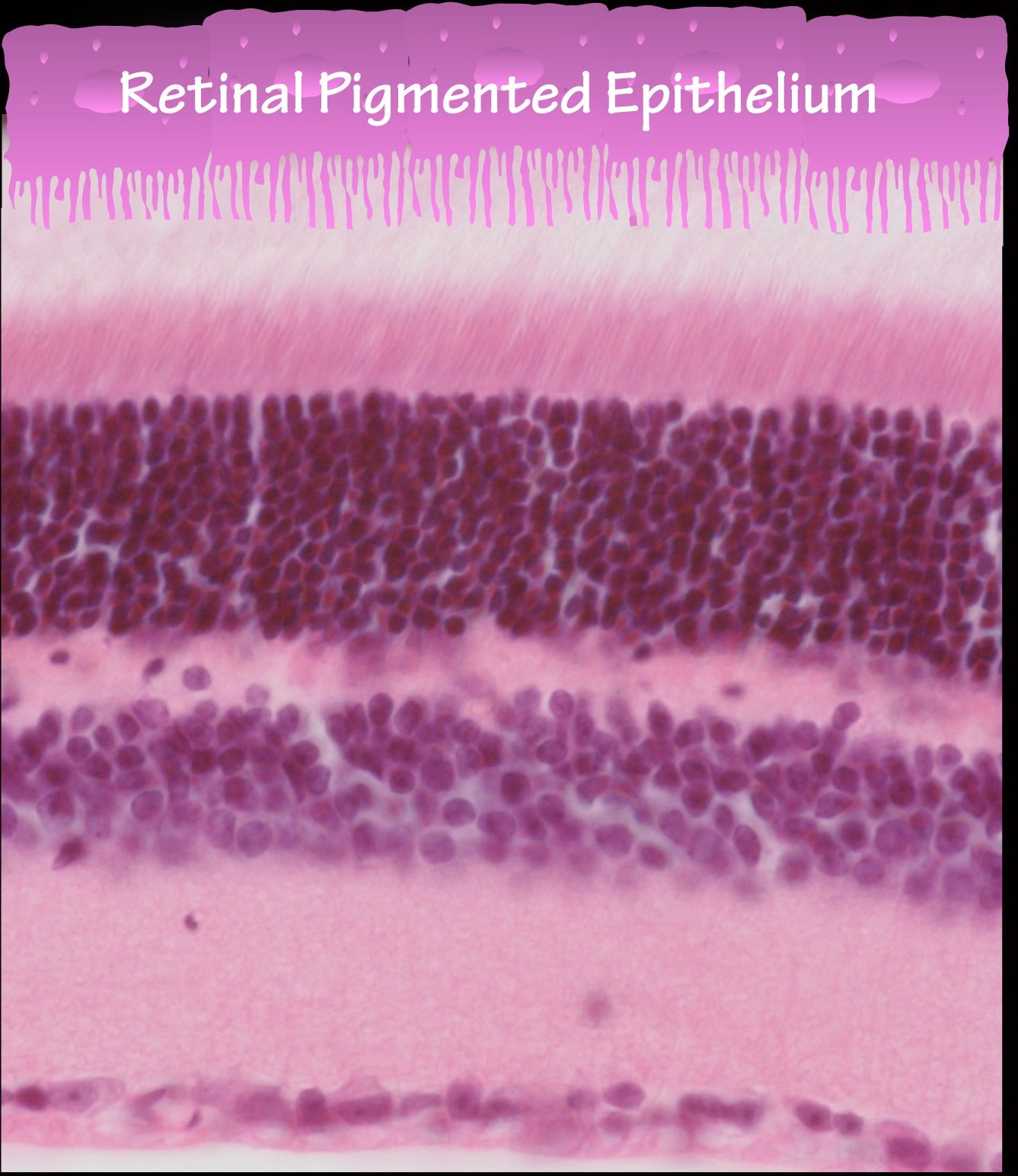
 The retina is comprised of three cellular layers, the outer nuclear layer, the inner nuclear layer and the ganglion cell layer. The outer nuclear layer contains only photoreceptors. Two types of photoreceptors are found in the vertebrate retina, rods and cones. Cone photoreceptors are responsible for vision during the brighter light intensities of the day and mediate color vision. Rod photoreceptors are 1000x more sensitive to light, and initiate vision in dim light. The light that is captured by PRs is converted into an electrical signal that is passed onto bipolar cells. Bipolar cells are located in the inner nuclear layer and pass the signal onto ganglion cells that are located in the ganglion cell layer. Ganglion cells are the output neurons of the retina that project to the brain. The other cell types found in the inner nuclear layer are amacrine cells, horizontal cells and Muller glia cells (Rollover image). Muller glia cells are the major glia cell type in the retina. They span the entire retinal thickness and play an important support role for the different retinal neurons. Muller glia cells are also part of the retinal-blood barrier. Amacrine and horizontal cells play an important role in modulating the primary signal from bipolar cells.
In the human eye, light is focused onto a small region of the retina, the fovea, where high acuity vision occurs. This area is highly adapted for daylight vision and contains only cone photoreceptors. Approximately 95% of photoreceptors in the rest of the retina are rods, with a central rod-rich ring that is almost free of cones. With the exception of the fovea, being absent in mouse, the human and mouse retina are very similar. Mice serve therefore as an excellent model to study retinal development and disease.
