The Circadian Clock: Brain Clocks & Circuits
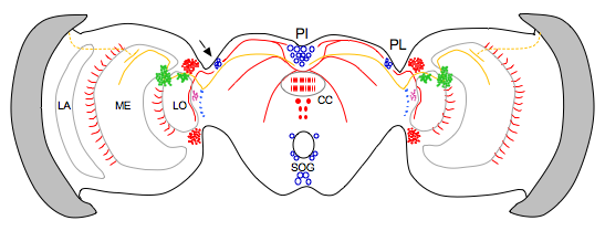
Using a strategy that relied on the coexpression of PERIOD (PER), TIMELESS (TIM), and CRY1, we identified four cells in the dorsolateral region of monarch butterfly brain (the pars lateralis, PL) as the putative location of circadian clocks (Sauman et al., 2005). Further study showed that CRY2 is also expressed in those cells, where it cycles in and out of the nucleus at the appropriate times to regulate the clockwork feedback loop (Zhu et al., 2008). Clock proteins are also expressed in large neurosecretory cells in the pars intercerebralis (PI); these cells may be part of a circadian network that contributes to circadian behaviors.
Importantly, the CRY proteins mark neural pathways that may be relevant for migration and circadian behaviors. CRY1-positive fibers connect the PL clock cells to axons from polarization-sensitive photoreceptors in the dorsolateral medulla. CRY1-positive fibers also connect the PL to the PI, which may be critical for the photoperiodic regulation of reproductive diapause and the initiation of the migratory state. A CRY2-staining neural pathway may connect the PL clocks or the PI to the central body region of the central complex, ultimately regulating circadian behaviors (e.g., daily flight activity, metabolic rhythms, and the sleep-wake cycle).

Brain clocks and circuits. Schematic representation of neurons and fibers expressing different circadian clock proteins in the monarch butterfly brain. Regions expressing TIM, PER, CRY1 and/or CRY2 are highlighted in blue. In these areas the four clock proteins partially colocalize. Areas expressing TIM or CRY1 are indicated in green. In these regions the two clock proteins do not colocalize. CRY1 positive fibers are represented by continuous orange lines. Projections of dorsal rim area photoreceptors are indicated by dotted orange lines. Neurons and fibers expressing exclusively CRY2 are represented in red and within the central body are shown as red circles and red hatching. Areas positive exclusively to TIM are indicated in light blue. Arrow, clock cell in the PL; PL, pars lateralis; PI, pars intercerebralis; SOG, subesophageal ganglion; CC, central complex; LO, lobula; ME, medulla; LA, lamina; RE, retina.