Nuclear Medicine Physics Imaging Research Interests
- Correction for causes of image degradation in nuclear medicine such as attenuation, distance-dependent spatial-resolution, and scatter.
- Detection, estimation, and correction of all forms of patient motion during imaging
- Tomographic image reconstruction for SPECT and PET.
- Combination of CT with SPECT and PET
- Design of the next generation of SPECT Imaging Systems
- Assessment of image quality by task performance studies using human and numerical observers.
- Quantification of activity and assessment of physiological function.
- Image segmentation and computer vision applications in nuclear medicine.
- Reduction in radiation dose to patients and personnel
- Applications of Deep Learning in Nuclear Medicine
One example of the research accomplishments of the laboratory is our work on the detection and compensation of patient motion during the up to 16 minute period which the patient is required to remain motionless for nuclear medicine studies. We have taken the approach of combining estimates of patient motion as determined from the nuclear medicine images themselves with information from our visual-tracking system (VTS) which tracks the motion of retro-reflective markers on stretchy bands wrapped about the patient to provide a combined correction of respiratory and bodily motion. Once motion is determined it is corrected by inclusion in iterative reconstruction as illustrated in the following figure. These investigations are funded by NIH grant R01-HL122484.

Clinical SPECT images showing benefits of respiratory motion correction (RM) as noted in top row versus second row which is without RM correction.
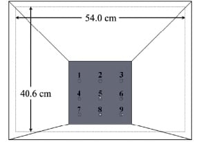
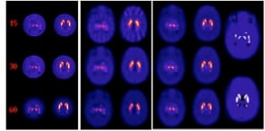
Another example of our current interests is the development of a multi-pinhole collimator (MPH) for use on one gamma-camera head of a general purpose SPECT system in combination with a fan-beam collimator on the second camera head for high resolution / sensitivity imaging of the striatal region of the brain in I-123 DaTscan imaging for Parkinson’s Disease. A rendering of the collimator is shown below followed by reconstructed simulated images of the strata for MPH, Fan, and Combined imaging. These investigations are funded by NIH grant R01-EB022092.
 |
| Rendering of the MPH collimator showing the aperture plate with 9 pinholes. |
 |
| A. Example XCAT transverse slice multi-pinhole (MPH) collimator noise-free reconstructions through substantia nigra (left) and the caudate and putamen (right) for 15, 30, and 60 angles for VOI over striata. B. Same for low-energy ultra-high-resolution (LEUHR) fan-beam only collimator reconstruction. C. Same for combined reconstruction using MPH and LEUHR fan-beam collimators. D. Matching slices through original XCAT DaTscan source distribution for substantia nigra (top), and caudate and putamen (bottom) with 8:1 striatal to rest of brain concentration ratio, and no activity in the ventricles. Note that separation of caudate and putamen can be seen in combined reconstruction. |
The third example of our current investigations is the development of a multi-detector-module multi-pinhole (MPH) SPECT brain-imaging system ideally suited for quantitative dynamic and high-spatial-resolution static SPECT imaging. Dynamic imaging will be enabled by obtaining sufficient angular sampling without the need for rotation. The system will automatically adapt its imaging characteristics (aperture size and number of pinholes open for imaging) in response to the imaging tasks and individual patients. It will thereby optimize lesion detection and quantification, as well as provide optimal data for pharmacokinetic analysis within structures throughout the brain. The prototype design for this system is illustrated in the following computer aided design (CAD) drawings. These investigations are funded by NIH grant R01-EB022521.
 |
 |
| Shown left is a frontal view and right is a view from behind of SolidWorks CAD renderings of the proposed prototype configuration of the 23 detectors of the system dedicated to brain SPECT imaging. Shown are detector modules with MPH aperture plates towards the brain and circular scintillation detector crystals opposed to them. | |


