Cool IR Case
17-year-old male with a long history of left-sided varicoceles status post surgical ligation in 2015 presents with recurrence of symptoms, referred to us for left gonadal vein embolization.
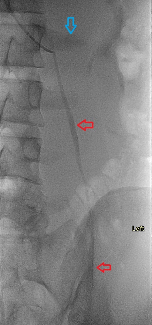
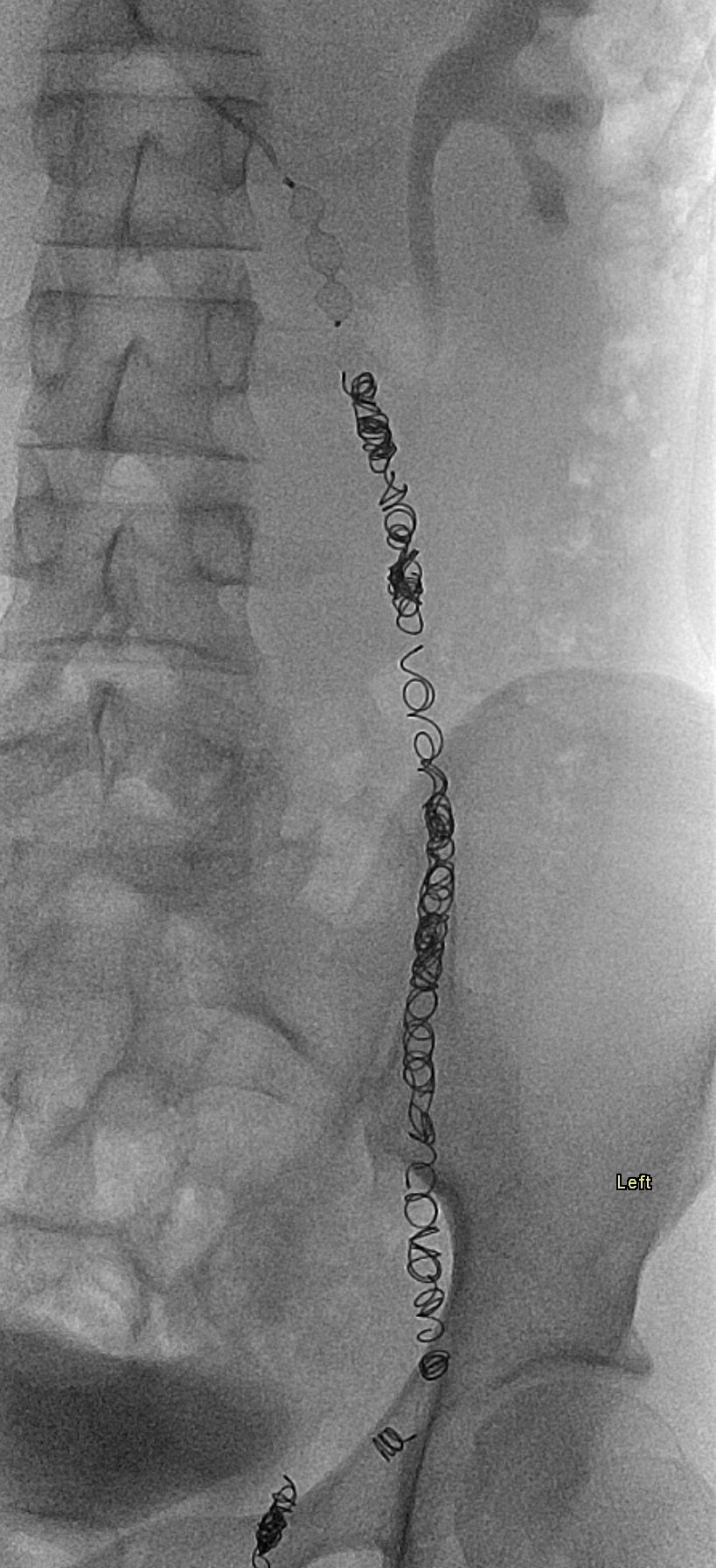
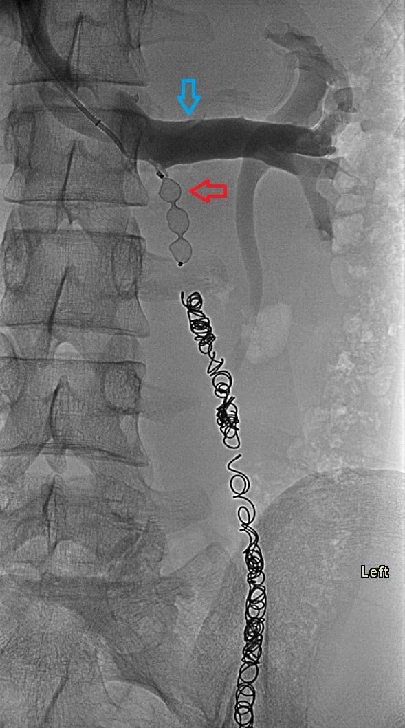
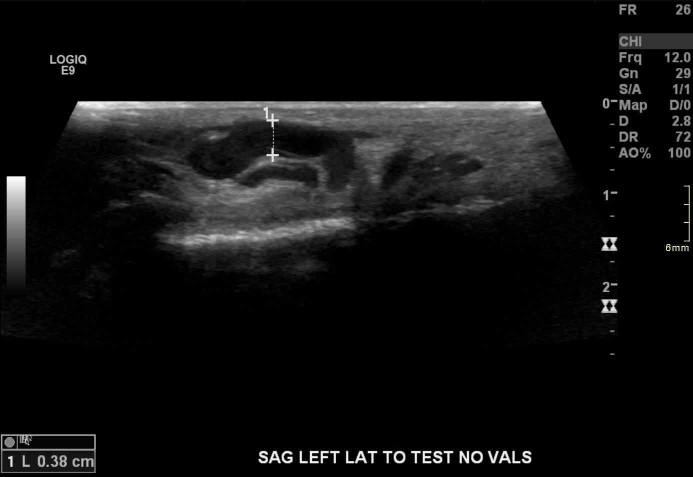
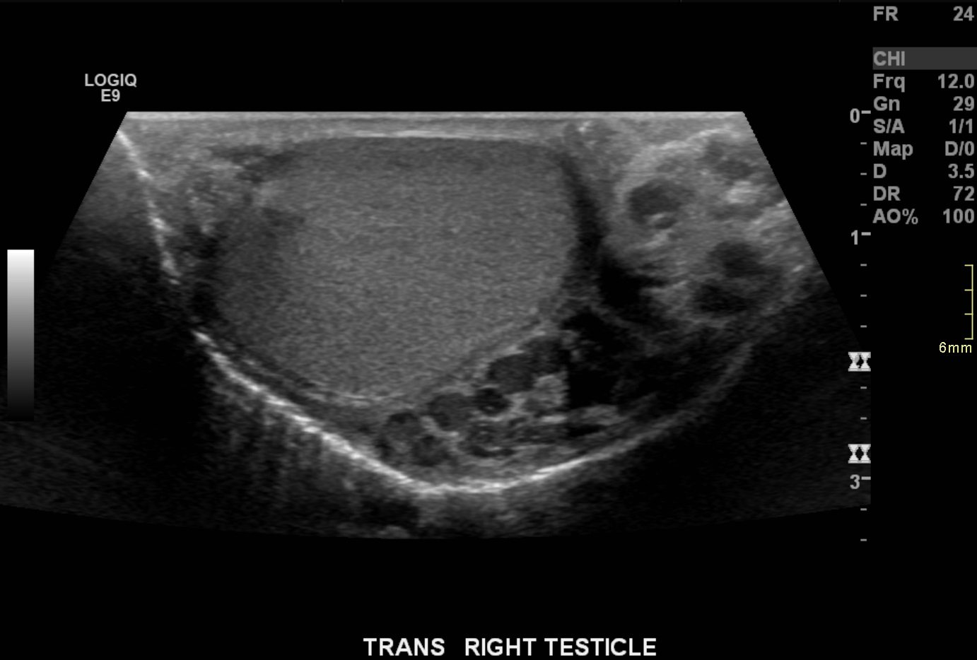
Ultrasound demonstrates enlarged veins (>3 mm) adjacent to the testicle. Angiogram demonstrates reflux of contrast into the left gonadal vein to the level of the scrotum, indicating venous valvular incompetence. Coil embolization and sotradecol sclerotherapy of the left gonadal vein were performed, with an Amplatzer plug at the top of the vein. Repeat angiogram demonstrates no contrast opacification of the vein. Red arrows indicate the left gonadal vein and the blue arrows indicate the left renal vein.
The patient's symptoms have resolved.
Aaron Harmon, MD, Assistant Professor, Interventional Radiology
 |
 |
 |
 |
||
 |
||


