Buscar Close Search
Buscar Close Search
brain synapse
Page Menu

Neuroscience Program

Neuroscience investigators focus on:

  • The neural, molecular and genetic mechanisms that underlie nervous system development and function, learning and memory, addiction, glial function, and circadian rhythmicity;
  • Mechanisms of synaptic neurotransmitter release, analysis of how neurotransmitter receptors and membrane channels operate, and how drugs act on these processes to modify cellular function and behavior;
  • Disorders of the central nervous system, with special emphasis on neurodegenerative disorders, amyotrophic lateral sclerosis, autism spectrum disorders, mental retardation and other developmental disabilities.
  • Development of new methods and therapies for neurological diseases, including disruption of mutant gene expression using chemically modified oligonucleotides, gene knock-down or replacement with adeno-associated viral vectors, and CRISPR-mediated gene correction.

The Graduate Program in Neuroscience brings together many components of the neuroscience community at UMass Chan Medical School.  Like the Graduate Program, the neuroscience community at UMass Chan Medical School is truly interdepartmental and interdisciplinary.  A critical and unique feature of the research environment at UMass Chan Medical School is that departmental affiliations affect letterheads but not interactions or collaborations. This atmosphere is especially conducive to the scientific growth of graduate students obtaining their degrees in an interdisciplinary field like neuroscience.

Participating faculty have primary appointments in 15 different departments, with the largest concentrations of faculty (> 10 each) located in the Departments of Neurobiology, Neurology and Psychiatry.  Clusters of neuroscientists are located in many other Departments, with 3 or more Program members in each of eight other departments:  Program in Molecular Medicine, Biochemistry & Molecular Biotechnology, Molecular, Cell & Cancer Biology, the RNA Therapeutics Institute, Microbiology, Radiology, Neurological Surgery and Pediatrics. This diversity of affiliations reflects the diversity of research interests in the Program, which range from investigation into basic mechanisms of neuronal function in model organisms and identifying novel disease genes to development of therapies for neurodegenerative diseases and improving clinical care for children with developmental disabilities.

Admission Requirements      Apply Now

REQUIREMENTS FOR SPECIALIZATION

Graduate students who specialize in Neuroscience will acquire a broad background in the concepts of contemporary neuroscience, gain exposure to state-of-the-art techniques and will acquire a foundation in the function of the nervous system through an integrated program of advanced coursework, laboratory research, and seminar and journal club attendance.

All graduate students within the BBS division of the Morningside Graduate School of Biomedical Sciences must complete the Biomedical Sciences Core Curriculum, consisting of Scientific Inquiry in Biomedical Research, Responsible Conduct of Research, Part 1 (Fall, Year 1), Preparation for Qualifying Exam (Fall, Year 2), and Responsible Conduct of Research, Part 2 (Year 3).  Students explore research areas of interest to them by participating in three rotations and then will select the faculty mentor who will supervise their thesis research.  Thesis Research Advisory Committee meetings are required annually during thesis research.  Students in the third year and beyond are also required to complete an annual Individual Development Plan, and the TRAC meeting will include discussion of progression toward both research and professional development goals.

In addition to the Morningside Graduate School of Biomedical Sciences Core Curriculum, students in the Graduate Program in Neuroscience are required to take at least three (3) graded elective courses of 2-4 credits each during their graduate career, of which one must be Cellular, Molecular and Developmental Neuroscience (BBS 780). This introductory course is usually taken in the Spring semester of the first year and covers topics including ionic mechanisms underlying neuronal excitability, neurosecretion, neurotransmitters and receptors, mechanisms of neuronal development and research methods in neuroscience. Two other elective courses are offered by the Program: Systems and Circuits Neuroscience (BBS 820) and Bases of Brain Disease (BBS 782). Elective courses offered by other graduate programs can also be taken to meet the elective course requirements. The elective courses are selected to yield a program of study tailored to meet the needs of each student.

Program in Neuroscience students are expected to attend the weekly Neuroscience Program Seminar Series lectures, featuring visiting experts from outside the university, and to participate in a seminar series in their home department. Students are also required to enroll in Neuroscience Seminar for two semesters. (Two discontinued courses, Communicating Neuroscience: Learning by Doing, and Journal Club in Neuroscience are treated as equivalent to Neuroscience Seminar in meeting this requirement).

View PhD Program Schedule  |   View courses

OUR LEADERSHIP & FACULTY

PROGRAM DIRECTOR

Dr. David WeaverDavid Weaver, PhD
Professor
email Dr. Weaver

OUR FACULTY

The Program in Neuroscience is interdepartmental, administered under the umbrella of the Department of Neurobiology. Participating faculty have primary appointments in several departments, with the largest concentration of faculty coming from the Departments of Neurobiology, Neurology, Psychiatry, Microbiology, Medicine, and Molecular Medicine.

View Affiliated Faculty

OUR STUDENTS

STUDENT EXPERIENCE

The program maintains a schedule of seminars and intramural research presentations that ensures a cohesive program. This atmosphere is especially conducive to the scientific growth of graduate students obtaining their degrees in neuroscience.

View current and past student listing

STUDENT SPOTLIGHT

Megan Fowler-Magaw, PhD candidate, Neuroscience

megan fowler magawMegan Fowler-Magaw studies a specific gene that is found in 97 percent of Amyotrophic lateral sclerosis (ALS) cases. The TDP-43 gene is a transactive response DNA-binding protein.

Learn more about Megan Fowler-Magaw

OUR STUDENTS IN THE NEWS

Getting Results…
  • Morningside Graduate School of Biomedical Sciences honors student achievement

    Morningside Graduate School of Biomedical Sciences honors student achievement

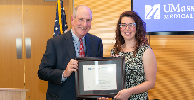
    In its annual pre-Commencement celebration, the Morningside Graduate School of Biomedical Sciences honored student scientists and recognized Mary Ellen Lane, PhD, for reaching her five-year mark as dean at the 2023 Student Achievement and Leadership Awards Ceremony.

    Read more
  • Morningside Graduate School of Biomedical Sciences speaker to urge pursuit of truth

    Morningside Graduate School of Biomedical Sciences speaker to urge pursuit of truth

    Geneticist Kathleen Morrill has been named speaker for the Morningside Graduate School of Biomedical Sciences Class of 2023. She will address her class during UMass Chan’s 50th Commencement on June 4.

    Read more
  • PhD candidate Kathleen Morrill receives Harold M. Weintraub Graduate Student Award

    PhD candidate Kathleen Morrill receives Harold M. Weintraub Graduate Student Award

    PhD candidate Kathleen Morrill received the 2023 Harold M. Weintraub Graduate Student Award for her work on canine behavioral genomics and its relevance to human genetics and psychiatric disorders.

    Read more

EXTERNAL AWARDS FOR RESEARCH TRAINING (CURRENT)

  • Allen.jpg

    Examining the Role of a Pathogenic HTT Isoform, HTT1a, in Somatic Expansion and RNA Aggregation in Huntington's Disease

    Huntington’s Disease – a devastating neurodegenerative condition – is caused by a defect in the Huntingtin gene, resulting in the production of alternative forms of Huntingtin mRNA and protein. This proposal will use small RNA drugs to reduce alternative forms of Huntingtin in mouse models of Huntington’s disease and determine the effect on disease features and outcomes. Findings from these studies will provide insight into the mechanisms underlying Huntington’s Disease to inform the development of future therapeutics.

    Read more
  • Liu.jpg

    Investigating the Role of cnb-1 and chpf-1 in GABA DD Motor Neuron Remodeling and Synapse Maintenance

    During development of the human brain, neurons are forming a mature neural circuit, which requires major rewiring of synaptic connections. Developmental remodeling helps the brain integrate rewired connections, which when disrupted is linked to neurological disorders such as schizophrenia and autism spectrum disorder. The goal of this project is to identify mechanisms regulating the highly conserved process of synapse remodeling. I will be using the simple model system Caenorhabditis elegans because it undergoes a striking example of remodeling in GABAergic dorsal D-class (DD) motor neurons. Prior work on synapse remodeling has largely focused on the presynaptic side, while my preliminary work has focused on the post-synaptic domain. The Francis lab has identified dve-1 to act as a transcription factor of remodeling, specifically synapse elimination. Through bulk RNA-sequencing (RNA-seq) I have identified 2 downregulated targets of dve-1, the calcineurin-like EF-hand protein CHP1/chpf-1 and the regulatory subunit of CalciNeurin, PPP3R1/cnb-1. Preliminary data has shown a defect of synapse remodeling in both cnb-1 and chpf-1. Additionally, I will determine the functional requirement and site of action for cnb-1 and identify the contribution of calcineurin phosphatase function and the proteasome to the effect of cnb-1 on synapse remodeling. In aim 2, I will further characterize the role of cnb-1 in both synapse elimination and maintenance, while also identifying the effect of calcium binding on synapse remodeling. In aim 3 I will identify potential targets of dve-1 in the remodeling pathway by using neuron-specific RNA-seq. My proposed work will advance the understanding of remodeling and the mechanisms of regulation, potentially providing targets for future exploration.

    Read more
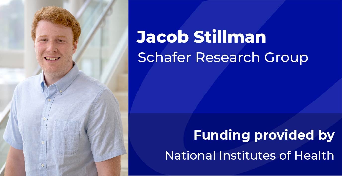
  • Stillman.jpg

    Investigating the Role of Endosomal Toll-Like Receptors in Remyelination

    Regulation of innate immunological self-tolerance, or the ability of cells to discern “self” from “non-self” has long been studied in the periphery in autoimmune disorders, especially in the context of nucleic acids (NA). Understanding of self-tolerance in the central nervous system (CNS), however, has not been thoroughly investigated despite expression of these NA-sensing TLRs by microglia, the primary phagocyte of the CNS. Published data from our lab highlights that microglia retain untranslated RNA transcripts from engulfed myelin for days after phagocytosis in vitro and in human multiple sclerosis patients. Based on these data, I hypothesized that these retained transcripts could aberrantly activate endosomal TLRs. I, thus, induced primary demyelination in UNC93B1 -/- mice, which lack functional NA-sensing TLRs, and observed that these mice remyelinate more efficiently than wildtype. These data suggest that signaling of NA-sensing TLRs suppresses remyelination during demyelinating disease. Several exciting questions have now arisen, which I will tackle in this proposal: 1) Is myelin phagocytosis causing aberrant endosomal TLR signaling? 2) Are microglia the primary cell type driving this response? 3) Does a specific NA-sensing TLR hinder remyelination? I hypothesize that TLR7 is aberrantly signaling in response to engulfed myelin RNAs in microglia and suppressing remyelination. To address these questions, I have acquired powerful in vivo molecular genetic tools to manipulate UNC93B1 and endosomal TLR function. I will first identify molecular pathways that are changed in microglia in vitro in response to chronic myelin phagocytosis and test whether these molecules are UNC93B1-dependent (Aim 1a). I will then determine if the UNC93B1 dependent effects that I observed on remyelination are microglia-specific (Aim 1b). Lastly, I will identify the endosomal TLR underlying these UNC93B1 effects (Aim 2). I am now in a strong position to molecularly dissect the role of NA sensing TLRs in remyelination during demyelinating disease, which has high long-term therapeutic potential.

    Read more
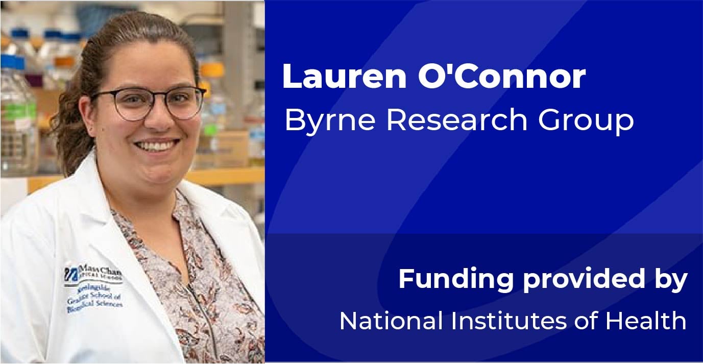
  • O'Connor L.jpg

    Investigating mechanisms of neurodegeneration

    Disease and injury cause a catastrophic loss of cognitive and motor abilities in the nervous system. Preventing neurodegeneration is critical to maintaining neuronal function. To develop effective treatments, we first need a greater understanding of the genetic and cellular mechanisms that determine whether the nervous system degenerates in response to various insults. Caenorhabditis elegans is a highly tractable model to dissect conserved molecular and cellular mechanisms. The goal of this proposal is to take advantage of the C. elegans model to identify regulatory mechanisms of axon degeneration. To reach this goal, I will apply my developing skills in detailed genetic analyses, laser axotomy, imaging, sequencing, and bioinformatics. The impact of this project is significant. In addition to providing a critical advance in understanding the fundamental mechanisms of neurodegeneration, it will also inform future therapeutic approaches that can be manipulated to protect the nervous system from degenerating.

    Read more

EXTERNAL AWARDS FOR RESEARCH TRAINING (PAST)

  • Gunner.jpg

    Dissecting ADAM10 function in microglia-mediated synapse elimination

    The goal of this proposal is to dissect the molecular signaling between microglia and neurons that regulates synapse elimination in response to changes in sensory experience. Despite compelling evidence that microglia, the resident brain macrophages, play important roles in eliminating synapses in development and disease, the precise neuron-to-microglia molecular signaling that drives this process is poorly understood. I recently discovered a signaling pathway necessary for microglia-mediated synapse elimination by utilizing the well-described circuitry of the mouse barrel cortex circuit as a model to manipulate sensory experience and dampen neuronal activity. Here I found microglia robustly engulf synapses in the barrel cortex following either whisker lesioning or trimming, and that this engulfment is dependent on the microglial CX3CR1 receptor and its canonical neuronal ligand, CX3CL1, but not complement. Using single-cell RNAseq I also found that neuronal Cx3cl1 was not differentially regulated in the cortex following whisker removal, but the protease Adam10, known to cleave membrane-bound CX3CL1 into a soluble form, is increased following lesioning. Importantly, pharmacological inhibition of ADAM10 resulted in synapse elimination defects that phenocopied CX3CR1 and CX3CL1-deficient mice. These data suggest that post-translational modification of neuronal CX3CL1 by ADAM10 is required to regulate microglial synapse elimination in the cortex following whisker removal. Several exciting new questions have now arisen, which I will tackle in this proposal: 1) What is the cellular source of ADAM10 and is it localized to synapses (Aim 1)? 2) Do other subcortical synapses within the barrel circuit remodel via ADAM10-CX3CL1-CX3CR1 signaling and does this differ between whisker lesioning and trimming (Aim 2)? I hypothesize ADAM10 is derived from layer IV excitatory neurons to regulate microglia- mediated synapse remodeling and that ADAM10 signaling is specific for cortical synapse rewiring after whisker trimming and lesioning, but not for sub-cortical synapse remodeling. To test this hypothesis, I have acquired powerful in vivo molecular genetic tools to manipulate ADAM10 function in specific cells. I have also developed collaborations to learn and perform cutting-edge whole tissue clearing by iDISCO to assess structural remodeling of entire circuits. Finally, I have a strong mentoring team that includes my mentor Dr. Dorothy Schafer with expertise in microglial function within neural circuits, my co-mentor Dr. Andrew Tapper with expertise in structural and functional mapping of brain circuits, and collaborators with expertise in iDISCO. Together, I am in a strong position to molecularly dissect how ADAM10 modulates neuron-microglia signaling necessary for remodeling brain circuits. This could be highly relevant for neurodegenerative disease where microglial dysfunction, synapse loss, and ADAM10 have been implicated. In the process, I will receive training in a variety of microscopy and molecular genetic approaches that will provide a foundation for my future career as an independent principle investigator at an academic institution focused on dissecting functions for glial cells within neural circuits.

    Read more
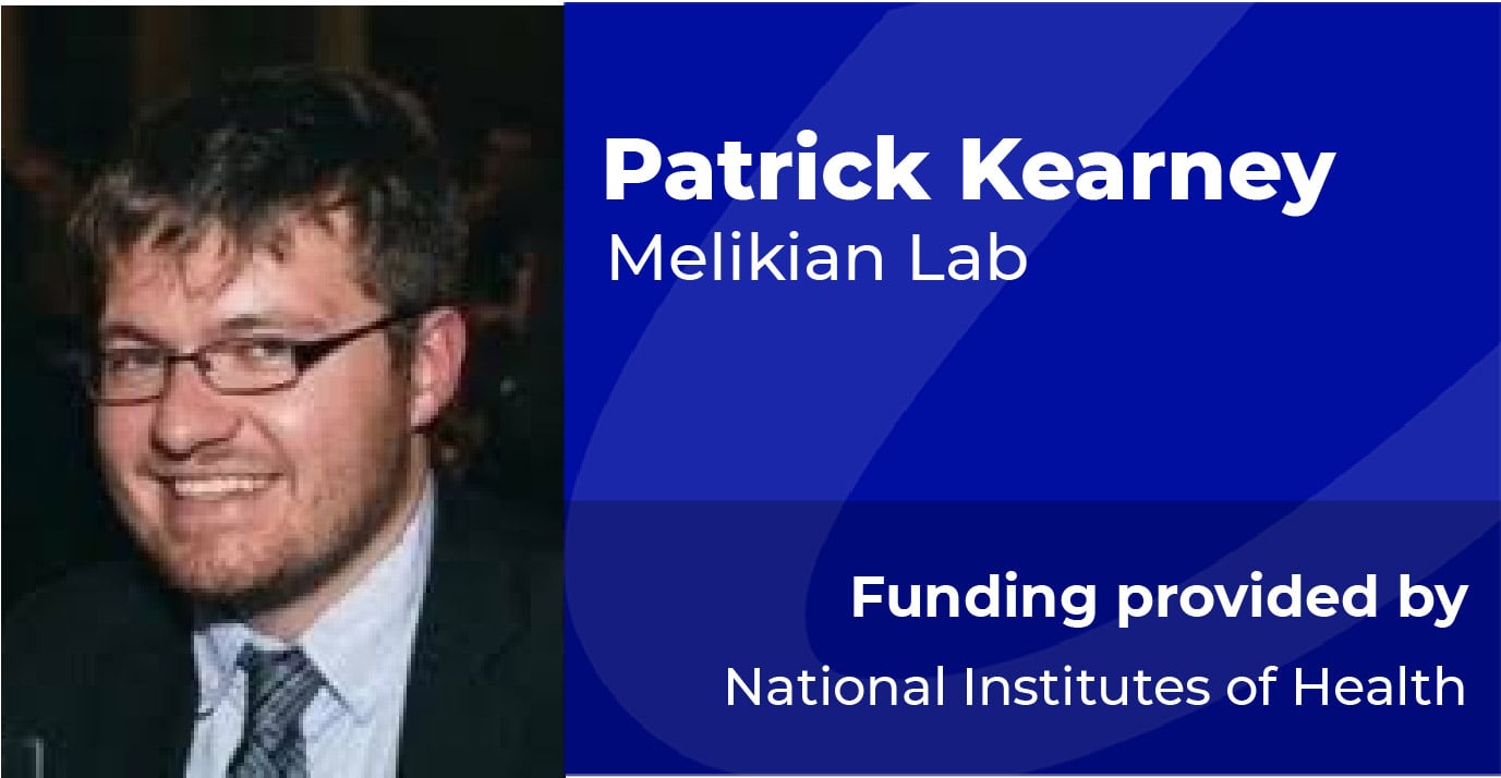
  • Kearney.jpg

    Gq Receptor Regulation of Striatal Dopamine Transporters

    Dopamine (DA) neurotransmission is vital for behaviors such as movement and reward, as well as, cognitive functions including mood, learning and memory. Several neuropsychiatric disorders are linked to alterations in DA signaling including Attention Deficit Hyperactivity Disorder (ADHD), schizophrenia, Parkinson's disease, and addiction. The DA transporter (DAT) is imperative for temporal and spatial control of DA signaling. DAT is located at the presynaptic terminal of DAergic neurons and facilitates the termination of DAergic transmission by rapidly clearing released DA. DAT is the primary target of addictive and therapeutic psychostimulants, which compete for DA binding and block uptake through the transporter, preventing DA clearance and leading to the hyper-locomotive and rewarding behaviors associated with drug use. Given that DAergic signaling is highly sensitive to DAT function, understanding the molecular mechanisms that control DAT function and availability is a critical missing piece of the puzzle in understanding DAergic neurotransmission and dysfunction in DA- related disorders. Over two decades of research support that DAT surface expression is acutely regulated by endocytic trafficking. Protein kinase C (PKC) activation with phorbol esters stimulates DAT internalization and thereby decreases DAT surface expression and function. Although considerable progress has been made to define the molecular mechanisms governing basal and PKC-regulated DAT trafficking, there are significant gaps in our understanding of this process in bona fide DAergic terminals. It is not clear how DAT is regulated in response to the endogenous presynaptic receptors that are activated upstream of PKC, such as Gq-coupled receptors, and how the complex signal events stemming from Gq receptor activation integrate to acutely control DAT surface expression. It is additionally unknown whether regulated DAT trafficking is region-specific, or whether altered DAT surface expression impacts DAergic signaling in the striatum. The proposed studies will leverage chemogenetic receptors to test how Gq activation impacts DAT surface levels in a cell- autonomous manner, in both dorsal and ventral striatum. We will capitalize on a novel conditional, inducible, in vivo gene silencing approach to determine the endocytic mechanisms that are required for Gq-mediated DAT trafficking, by both chemogenetic and endogenous presynaptic receptors. We will further employ ex vivo fast- scan cyclic voltammetry to investigate how presynaptic DAT trafficking impacts DA signaling. I anticipate that at the completion of these studies, we will have gained a more in-depth understanding of the complex mechanisms underlying DAT regulation at presynaptic DAergic terminals, and its potential influence on synaptic DA homeostasis.

    Read more
  • Gutierrez.jpg

    Fluorescent visualization of complement-dependent pannexin activity in microglia

    The goal of this project is fluorescently visualize ATP release and extracellular accumulation at the surface of stimulated microglia. The development of this innovative technology has the potential to enable spatiotemporal imaging of microglial extracellular signaling. For this project, I am exploiting the presence of the cell's glycocalyx to attach ATP-sensitive biosensors at the sites of ATP accumulation. There are two aims to this project: 1) to synthesize a novel, polyhistidine binding moiety that covalently modifies the glycocalyces of living cells and binds recombinant biosensors to measure ion and metabolite efflux and accumulation; 2) to visualize and measure ATP release from pannexin channels in C5a stimulated microglia. The completion of these aims will yield a transformative set of chemical-biological tools and methodologies to investigate the physiology and pathophysiology of pannexin-dependent activity in glia, and potentially in living animals.

    Read more