Page Menu
Biochemistry and Molecular Biotechnology Program
The Graduate Program in Biochemistry and Molecular Biotechnology offers graduate study and research focused in the areas of molecular, cellular and regulatory biochemistry, molecular biophysics, chemical biology, and structural biology. Students receive a rigorous foundation in modern biomedical science through an integrated program of laboratory research, advanced coursework, and attendance and participation in seminar programs. Students also organize and participate in a weekly informal seminar series in which they present recent research results.
Specific areas addressed within program laboratories include protein structure, function and evolution, regulation of gene expression, chromatin structure and epigenetics, translational regulation, membrane transport, ion channel function, drug resistance, cell cycle control, DNA replication, and neurodegenerative disease.
Admission Requirements Apply Now
REQUIREMENTS FOR SPECIALIZATION
All Basic Biomedical Science students must complete the core curriculum as well as electives required by their program. Students in the Biochemistry and Molecular Biotechnology (BMB) program must take 3 graded elective courses of 2-4 credits each, two of which must be part of the BMB program course curriculum. The third elective is typically taken in Year 2; however, the student should enroll in a course (regardless of when it is offered) that is most relevant to their graduate research. The plan of coursework is designed to be flexible in order to accommodate each student’s needs and areas of interest.
All students in thesis research are required to give an annual research presentation to the Department in a seminar series that runs from September through May each academic year.
View PhD Program Schedule | View Courses
FACULTY
Our faculty include, Howard Hughes Medical Institute Investigators, W. M. Keck Foundation Distinguished Young Scholars, Burroughs Wellcome Fellows, Worcester Foundation for Biomedical Research Scholars, a Pew Scholar and a faculty member who patented discoveries in RNAi.
Research areas of our faculty include:
- Biophysics
- Chemical Biology
- Computational Biology
- Cell & Developmental Biology
- DNA/RNA & Epigenetics
- Human Disease & Therapeutics
- Membrane Biology
- Neurobiology
- Structural Biology
View the affiliated faculty listing for the Biochemistry and Molecular Biotechnology Program.
The Graduate Program in Biochemistry and Molecular Biotechnology is an integral part of the Department of Biochemistry and Molecular Biotechnology. Our graduate students are core members of our world-class research teams and a central part of our departmental culture. Students organize our weekly departmental research seminar and happy hour, where they and other department members present their research. They also present at our annual departmental research retreat, the university research retreat and at national and international meetings. Many of our students are funded by NIH pre-doctoral fellowships and several have been awarded the prestigious Harold Weintraub award.
EXTERNAL AWARDS FOR RESEARCH TRAINING (CURRENT)
-
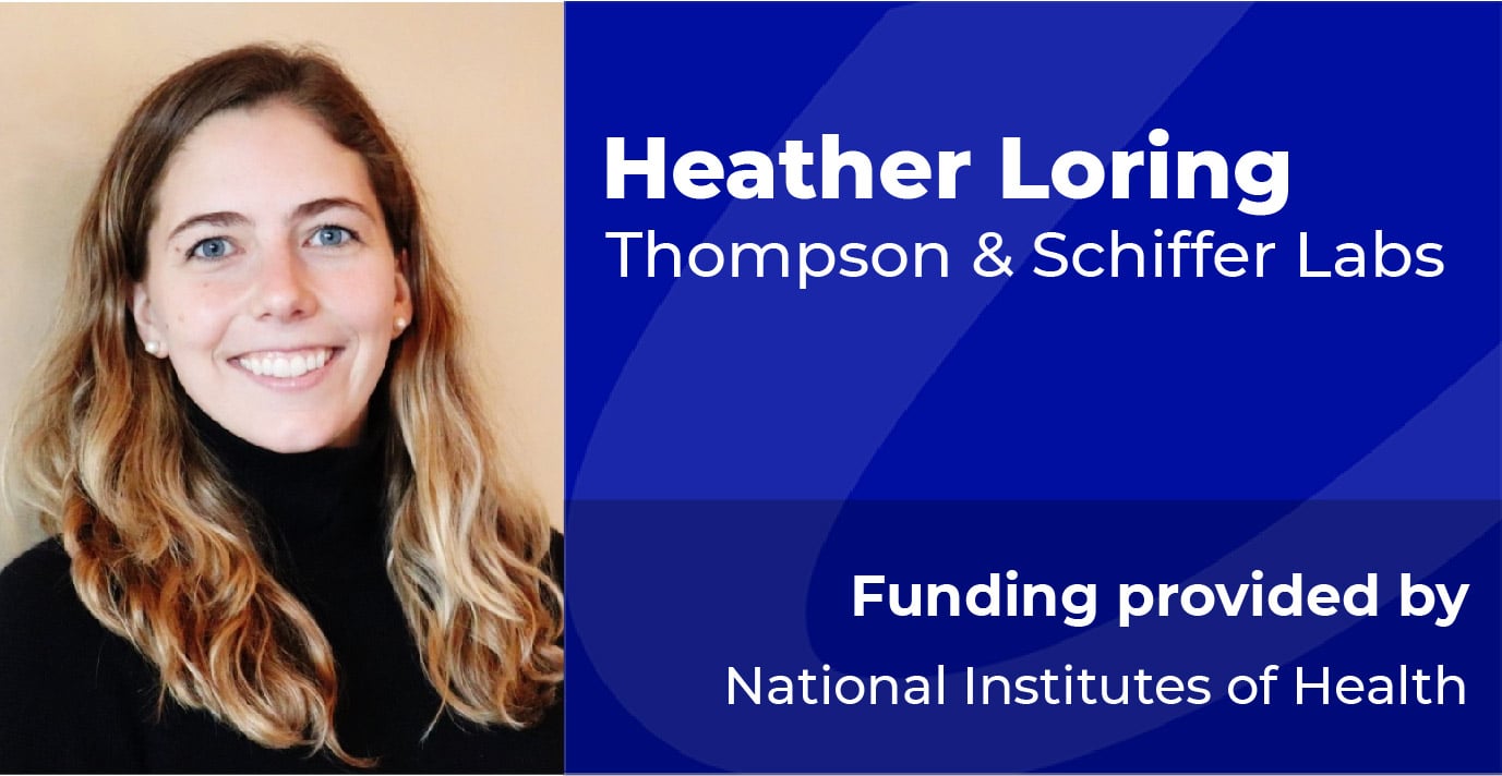
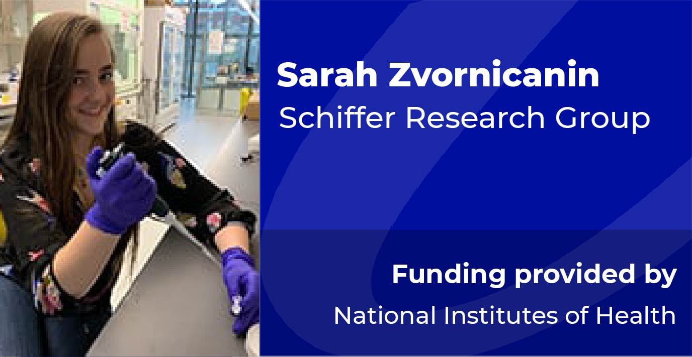
Human T-cell leukemia virus type-1 (HTLV-1) is an oncogenic human retrovirus affecting over 20 million people worldwide. HTLV-1 infection can cause adult T-cell lymphoma (ATL) and other serious inflammatory diseases. Estimates report that 5-10% of HTLV-1 infected patients will develop a serious condition such as ATL, which has poor 4-year survival and high relapse rates. HTLV-1 has persistent infection rates across the globe and reaches up to 45% prevalence in certain communities. Despite this impact on human health, there are no direct-acting antivirals (DAAs) or vaccines against HTLV-1. HIV-1 and HTLV-1 are from the same viral family and encode for a homodimeric aspartyl protease crucial for cleavage of functional proteins from viral polyproteins. The activity of HIV-1 and HTLV-1 protease is essential to their viral life cycles. The Schiffer laboratory has extensive experience with viral protease crystallography and inhibition, especially with viral proteases for HIV-1, HCV NS3/4A, and SARS-CoV-2 main protease. This expertise uniquely positions me to design, synthesize, and characterize potent, resistance-thwarting protease inhibitors against HTLV-1 protease. Resistance-preventing DAA design is essential because of the selective pressure applied during DAA treatment. An ideal and proven strategy for developing a highly potent and resistance-preventing viral protease inhibitor is to target the active site through rational design using the substrate envelope. The substrate envelope for HTLV-1 protease has not been characterized and we lack a detailed understanding of the protease substrate specificity. I hypothesize that by translating strategies from our design of HIV-1 protease inhibitors, namely characterizing HTLV-1 protease’s substrate specificity, I can design potent and resistance-preventing DAAs for HTLV-1 protease. Aim 1: Characterize the structural basis for HTLV-1 protease substrate specificity. HTLV-1 protease cleaves six substrates by recognizing cleavage sites between individual proteins of the viral polyprotein. I will investigate the molecular basis of this recognition underlying protease specificity by determining cocrystal structures of the protease with bound substrates. The conserved volume inhabited by the substrates will define the substrate envelope and inform inhibitor design for HTLV-1 protease. Aim 2: Rationally design, synthesize, and characterize inhibitors of HTLV-1 protease to optimize potency. HIV-1 and HTLV-1 proteases share an active site amino acid sequence identity of 45% and high structural similarity. Therefore, I will begin inhibitor design by testing a selection of our in-house HIV-1 protease inhibitors, which have already shown low (1 µM) to moderate (30 nM) potency against HTLV-1 protease. I will combine experimental inhibition assays with cocrystal structure analysis to identify lead compounds for inhibitor design. I will leverage substrate specificity of the protease by moving inhibitor design towards compounds that mimic the shape of substrates, leveraging the substrate envelope (Aim 1), and the interactions between protease and substrate. I aim to produce novel, highly potent (sub-nM) inhibitors that will be promising DAAs for further investigation against HTLV-1.
Read more
EXTERNAL AWARDS FOR RESEARCH TRAINING (PAST)
-
After injury, axons begin to die via a process that is characterized by axonal fragmentation and disintegration of myelin sheath. This process is often termed Wallerian degeneration after Augustus Waller. Wallerian-like degeneration, which is morphologically similar to Wallerian degeneration, is associated with the early stages of many neurodegenerative diseases, including as Alzheimer’s, Huntington’s, and Parkinson’s Diseases. Wallerian degeneration was long thought to occur passively, but the discovery of proteins that actively prevent or promote degeneration negated this idea.
Read more
-
All cells must replicate their genome once per cell cycle. To ensure proper duplication, cells integrate hundreds of factors that copy, surveil, and repair our genetic information. Proliferating Cell Nuclear Antigen [PCNA] and Rad9-Rad1-Hus1 [9-1-1] are ring-shaped clamps that act as master “conductors” that regulate many of the factors that replicate and maintain our DNA. PCNA is a homotrimeric ring that coordinates the replisome during DNA synthesis to work in tandem with DNA repair, chromatin remodeling, and cell cycle progression. When cells experience dsDNA breaks, they use the heterotrimeric clamp 9-1-1 to coordinate specific “SOS” repair factors. The collaborative efforts of both clamps are critical for genome stability. Many cancers are linked to inappropriate clamp coordination and changes in their expression. Because sliding clamps are central to many oncogenic pathways, we must address how they regulate themselves and their client partners. This proposal aims to address the following questions about sliding clamps: 1) How do sliding clamps coordinate their various partners? 2) Does the time sliding clamps spend on DNA influence genome stability? and 3) What determines site-specific loading of sliding clamps? I propose a multidisciplinary approach to address these questions about sliding clamps by investigating two-disease causing PCNA variants [PCNA-S228I [serine to isoleucine] and PCNA-C148S [cysteine to serine]] and the loading mechanism of 9-1-1. I hypothesize that sliding clamps control genome integrity via site-specific loading, proper partner interactions, and residence-time on DNA. I further hypothesize that PCNA-S228I and PCNA-C148S disrupt genome integrity by either promoting premature DNA dissociation or disrupting partner interactions. Finally, I hypothesize that the Rad17 subunit alters the clamp loader structure to specifically load the 9-1-1 clamp at sites of DNA damage. In aims 1 and 2, I will use PCNA-S228I and PCNA-C148S to address how clamps “choose” their partners and regulate their time on DNA. I will use x-ray crystallography, unfolding experiments, and a series of functional assays to determine how each variant compromises genome stability. In aim 3, I will determine the loading mechanism of clamp 9-1-1 to address how clamps are loaded to specific sites in the genome. I will use cryo-electron microscopy to determine how Rad17-RFC binds to clamp 9-1-1. Collectively, my work will broaden our insight into the factors that cause genome instability which may augment the development of personalized chemotherapeutics.
Read more
-
The apolipoprotein-B mRNA-editing catalytic polypeptide-like 3 (APOBEC3, or A3) family of cytosine deaminase enzymes forms part of the innate immune system - hypermutating cytosine (C) to uracil (U) in single-stranded DNA (ssDNA), as a first response to invading viruses. Specific A3 enzymes, including A3G, were first shown to potently restrict human immunodeficiency virus (HIV). However, expression of these enzymes is a delicate balance – low level deamination of the HIV genome provides a constant source of viral mutation, contributing to high viral fitness and rapid resistance to anti-viral drugs. Moreover, upregulation of other A3 enzymes, like A3A and A3B, is linked to many cancers, helping to expand genetic diversity in tumors to supercharge progression, metastasis, recurrence, and drug resistance. Abolishing activity of specific A3 enzymes may slow viral evolution and prevent tumor recurrence.
Structural examination of A3 active sites reveal their close homology to that of human cytidine deaminase (CDA), which converts C to U in single nucleosides. Introducing CDA inhibitors (transition state analogues, or TSAs) in place of the target C in ssDNA enables delivery to the A3 active site. Yet, development of selective inhibitors against individual A3 family members remains a challenge. Recently, our lab solved the co-crystal structures for A3A-ssDNA and A3G-ssDNA, and have also discovered that both the tertiary structure and DNA sequence flanking the target cytosine confer specificity to different A3 enzymes. I hypothesize that by exploiting these preferences – through manipulation of DNA sequence, oligonucleotide tertiary structure, and rational placement of chemical modifications – I can create potent and selective A3 inhibitors. Leveraging the Watts lab’s expertise in nucleic acid chemistry and the Schiffer lab’s extensive experience with A3G, I will first develop A3G-selective inhibitors, and then A3A and A3B inhibitors.
Read more
-
As the most abundant and deadliest entities on earth, bacteriophage play an essential role in many biological environments. While there are well-developed phage model systems that have informed our understanding of phage in the past 60 years, these systems have structural limitations. Here, we use a unique thermophilic siphovirus, P74-26, with extraordinary strength and stability to fill in the knowledge gaps that remain and take a closer look at some of the fascinating abilities of a phage that developed under the evolutionary pressure of a hot spring. Double-stranded DNA (dsDNA) viruses, which include bacteriophage along with herpesviruses, adenoviruses, use a powerful ATPase motor to pump their genome into an immature structure called the procapsid. Genome loading leads to immense internal pressure, resulting in a conformational change from a spherical particle to an expanded, pressurized icosahedron. The extreme stability of P74-26 despite high temperature and pressure makes it a novel tool for elucidating the intricacies of phage assembly and thermodynamics. In this study, we will combine cryo-EM, SEC-multi angle light scattering, and mass spectrometry to examine physical and mechanistic aspects of P74-26. A structure of the uniquely long phage tail tube will provide perspective into the mechanism of DNA ejection and possible evolutionary advantages for such length. Additionally, we have found that the major capsid protein spontaneously assembles, allowing us to create a controlled system for determining the essential components for viral head stability.
Read more
-
A leading cause of Cystic Fibrosis (CF) is premature termination codons (PTCs) in the cystic fibrosis transmembrane conductance regulator (CFTR) gene. Suppression of translation termination at PTCs—i.e. PTC readthrough—to restore full-length CFTR protein may be a treatment strategy. Yet, current PTC readthrough drug candidates for CF are toxic (e.g. aminoglycosides) or ineffective (e.g. ataluren). Efficacy of PTC readthrough depends on efficiency of translation termination at the PTC. Thus, manipulating the molecular mechanisms of CFTR PTC termination to lower efficiency may improve PTC readthrough efficacy. However, strategies for such manipulations are limited in the absence of a detailed understanding of translation termination on CFTR PTCs. In the current model for normal termination, eukaryotic Release Factors 1 and 3 form a complex (eRF1•eRF3) that releases a newly synthesized protein from the ribosome. eRF1 recognizes a tetra-nucleotide stop codon at the end of an open reading frame, and catalyzes peptidyl-tRNA hydrolysis. Poly-A binding protein (PABP), which binds at 3′ ends of mRNA, recruits eRF3 and enhances termination efficiency. However, it remains unclear how PABP, eRF1, eRF3, and the tetra-nucleotide stop codon recognize the PTC to produce truncated CFTR protein. The goal of this proposal is to determine the biochemical and structural mechanism of translation termination. With guidance from Dr. Andrei Korostelev (expert in biochemical and structural mechanisms of translation), Dr. Allan Jacobson (expert in premature translation termination and PTC read-through), Dr. Phillip Zamore (RNA biochemist), Dr. Chen Xu (cryo-EM instrumentalist), and Dr. Nikolaus Grigorieff (expert in cryo-EM method development), release assays will be optimized to study the efficiency of translation termination mediated by eukaryotic release factors, and ensemble time-resolved (ENTIRE) cryo-EM will be used to capture structural intermediates of enzymatic reactions. Aim 1 will use defined mammalian translation systems to measure the individual effects of stop codon context, eRF1•eRF3, and PABP on the termination efficiencies (kcat/KM) of CFTR PTCs and the true stop codon. Aim 2 will visualize how the ribosome terminates on CFTR PTC G542X in its natural sequence context using ENTIRE cryo-EM. Collecting data at multiple time points will identify conformational changes and interactions between mRNA sequence, eRF1•eRF3, and PABP during termination. To reveal the termination mechanism on CFTR PTCs, structures and their progression intermediates will be compared with those recorded on the true CFTR stop codon. If successful, this study will reveal key molecular determinants of CFTR PTC termination, and may inform strategies to induce PTC readthrough for CF treatment.
Read more
-
The novel NAD glycohydrolase, SARM1, is an active executioner in progressive axonal and neuronal degeneration1. This type of degeneration, termed Wallerian degeneration, defines a number of diseases, including neuropathies, traumatic brain injury and neurodegenerative diseases, yet no therapies exist. In fact, prior to the discovery of SARM1’s role in triggering Wallerian degeneration, the process was believed to occur passively.
Read more
POST-DEGREE CAREERS
Biochemistry and Molecular Biotechnology graduates pursue a variety of career options. Many go on to postdoctoral training and subsequent academic careers. Others pursue opportunities in the pharmaceutical industry and biotech, with recent graduates taking positions at Vertex, Biogen, Pfizer, Moderna, and Genzyme, among others. Biochemistry and Molecular Biotechnology graduates have also pursued diverse career paths, such as law, publishing and policy.
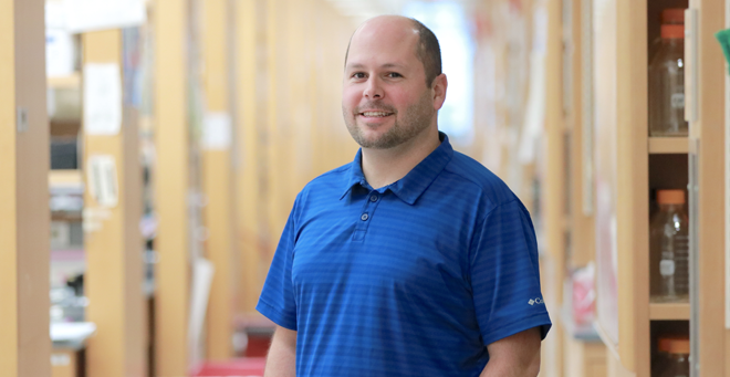 Brian Kelch, PhD
 Read more