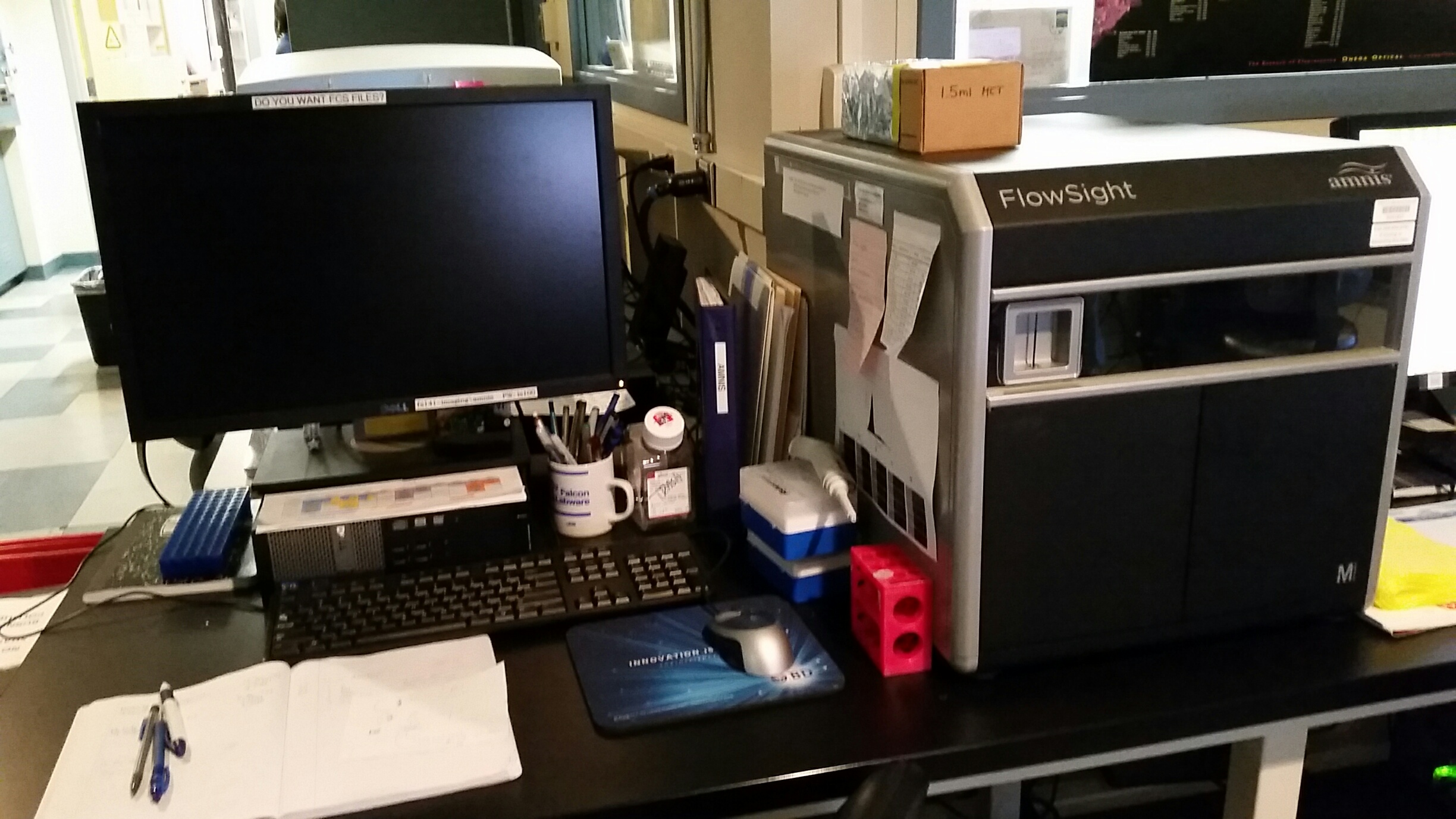
Core Amnis Flowsight Imaging Cytometer


Flowsight Imaging Channel Guide
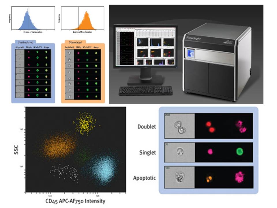
This unique cytometer quantitatively detects brightfield, darkfield and fluorescent images with high sensitivity CCD cameras. The fluorescence amplification and minimal noise detection results in very high signal sensitivity. In addition to being able to see the cells displayed in dot plots and histograms, the imaging provides analysis tools not available in conventional cytometers such as texture and spot counting. In the photo above, the color dot plot is a 5-way differential of cells in whole blood using only CD45 fluorescence and darkfield side-scatter. The Flowsight is equipped with violet, blue and red lasers for fluorescence detection in up to 10 channels. Running this instrument is straightforward requiring only laser power adjustment. Make an appointment and core facility personnel will run your samples while showing you how to use the instrument independently.
