MR Imaging of enzyme activity (Molecular Sensing Research)
Our lab is currently developing imaging probes that can be used across an array of imaging methods (MRI, SPECT, photoacustic (PA) imaging) to detect specific molecules and disturbances in these molecules in the initial phase of disease development.
MR Signal Amplification for Receptor Imaging
We have developed a novel class of MR detectable sensing probes, i.e. chemical compounds that respond to the presence of a given molecule (enzymes and various low-molecular weight analytes) that can induce a specific change in water relaxation, thereby producing visible changes in MR signal intensities on T1 weighted MR images.
One such imaging probe is di-HT-DTPAGd, a di-5-hydroxytryptamide of a commercially available gadolinium-based MR contrast agent, DTPA(Gd). In the presence of endogenous peroxidases (enzymes which become elevated during the course of certain diseases), individual molecules of di-HT-DTPAGd will polymerize and are then retained at the site of inflammation. The increase in MR signal (MRamp) which follows results from both an increase in the concentration of the paramagnetic contrast agent at the site of the enzyme, and from an inherent change in the relaxivity, i.e. the ability to shorten relaxation times of water protons. di-HT-DTPAGd is now available for investigational research on a multigram scale. We have also developed macrocyclic and open-chelate types of oxidoreductase-specific paramagnetic substrates and are also working on macrocyclic chelates of manganese cations, which like gadolinium, are paramagnetic.
The use of “enzyme sensing” MR contrast agents is expected to have wide-spread utility for in vitro high throughput screening as well as for in vivo detection of antigen expression patterns. The inherent chemical customization of these probes makes them ideal platforms for building new theranostic compounds.
Our laboratory is focusing on two major applications:
- Imaging of myeloperoxidase activity in cardiovascular disease and cancer.
- Targeted imaging of cancer-specific molecules using MRamp technique.
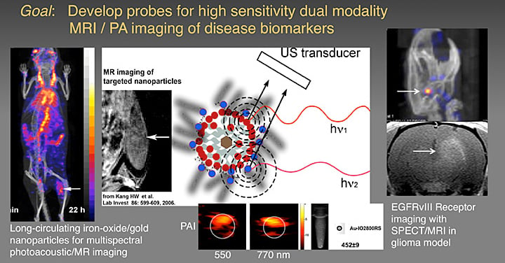
 A) sagittal and B) axial SPECT images showing accumulation of radionuclide- labeled antibody conjugates in EGF receptor-expressing tumors. Arrows indicate position of the tumor. C) sequential MRI brain images of EGF receptor-expressing tumors after the injection of a peroxidase specific imaging probe in vivo; (–) temporal washout of imaging probe with no pre-injection of targeted peroxidase enzyme- anti-EGF receptor antibody conjugates; (+) washout of the same probe following the injection with anti-EGF receptor conjugates in the same MRI matched slices. © ISMRM
We are also developing probes for high sensitivity dual modality MRI/PA imaging of disease biomarkers.
