Imaging in CVA Models - Detection of brain aneurysm instability using molecular imaging
There is now evidence that local and sustained inflammation is an initial component of a variety of disease such as cardiovascular disease, diabetes, and dementia. In many cases large numbers of neutrophils carrying primary myeloperoxidase (MPO)-rich granules are the first responders to local tissue perturbations resulting in a downstream infiltration of monocytes and signaling molecules which exacerbate inflammation and tissue damage.
Human Brain Aneurysm Rupture Risk Assessment Using Non-invasive Imaging
Goal: Develop molecular imaging markers to detect aneurysm instability thus allowing patient stratification for treatment
Cerebrovascular accidents are a leading cause of morbidity and mortality in the United States. Hemorrhagic stroke, secondary to intracranial aneurysm rupture, is a devastating medical emergency. There is currently no objective diagnostic means to identify aneurysms that are at high risk for rupture despite significant improvements in treatment. In addition, aneurysm rupture often occurs with no warning signs (unlike ischemic stroke which is often preceded by a transient ischemic attack). Thus there is an urgent need to develop new diagnostic methods that can quickly and safely identify patients at high risk for aneurysm rupture.
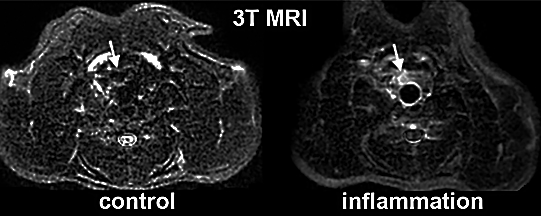
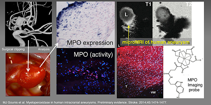
While the exact events leading to aneurysm rupture have yet to be fully elucidated, there is evidence suggesting that local inflammation may play a major role in aneurysm instability. Recent animal and human studies have indicated that one inflammatory enzyme,myeloperoxidase (MPO), is closely linked to aneurysm rupture. We have developed a technique, MRamp (magnetic resonance signal amplification), that allows MR detection and imaging of anatomically localized MPO based on the localization and retention of enzyme-sensitive MR substrates at sites of vascular inflammation. MRamp has been successfully used to image local MPO activity in an experimental animal model of aneurysm inflammation following injection of pro-inflammatory mediators.
Animal Studies
 Control and inflamed model aneurysms imaged using 3T MRI after the administration of an MPO-specific imaging probe di-HT-DTPAGd.RSNA©
Human Studies

MJ Gounis et al. Myeloperoxidase in human intracranial aneurysms: Preliminary evidence. Stroke 2014; 45:1474-1477.
Tissue from human brain aneurysms was harvested after clipping (3 samples from ruptured aneurysms and 20 from un-ruptured aneurysms (UIA)) and were histologically and biochemically evaluated for the presence of myeloperoxidase. All ruptured aneurysms were myeloperoxidase positive. Of the UIAs, half were myeloperoxidase positive. For each UIA, its 5-year aneurysm rupture risk was determined to be higher for myeloperoxidase-positive UIA (2.28%) than myeloperoxidasenegative UIA (0.69%), and the distributions were statistically different (P<0.005, Wilcoxon–Mann–Whitney test). The likelihood for myeloperoxidase-positive UIA was significantly associated (P=0.031) with aneurysm rupture risk (odds ratio, 4.79; 95% confidence limits, 1.15–19.96) suggesting that myeloperoxidase may potentially be used as an imaging biomarker of aneurysm instability.