Research
About the UMass Chan Medical School Cryo-EM Facility
The Massachusetts Facility for High-Resolution Cryo-Electron Microscopy at UMass Chan Medical School was opened on Tuesday, Oct. 4, 2016. The first-of-its-kind facility in New England opened new windows into the world of biology with the ability to see single molecules and their dynamics and macromolecular assemblies inside cells in unprecedented detail.
Video: Cryo-Electron Microscopy facility opens
What is Cryo-EM?
Cryo-EM -- Cryo-Electron Microscopy is a technique used in structural biology. It uses high speed electrons as source beam to obtain structural information of the biological system studied. Usually, the biological sample is embedded in vitrious ice by rapid freezing. The cryo sample is then observed and imaged in an electron microscope under liquid nitrogen temperature.
If you are new to Cryo-EM, you might find this short youtube video made by Gabriel Lander useful.
Cryo-EM Specimen Preparation Pipeline
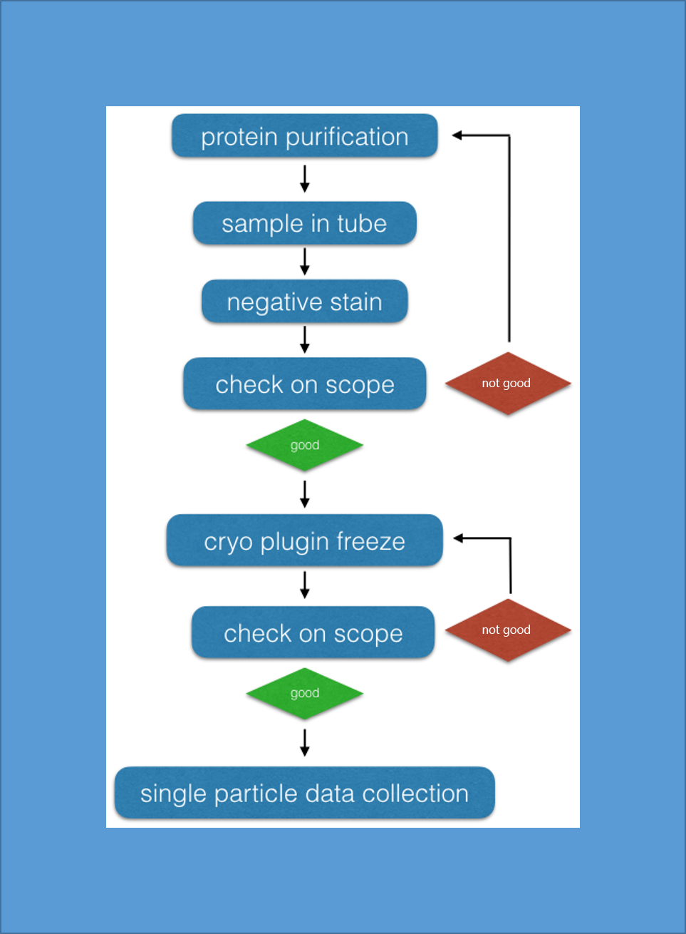
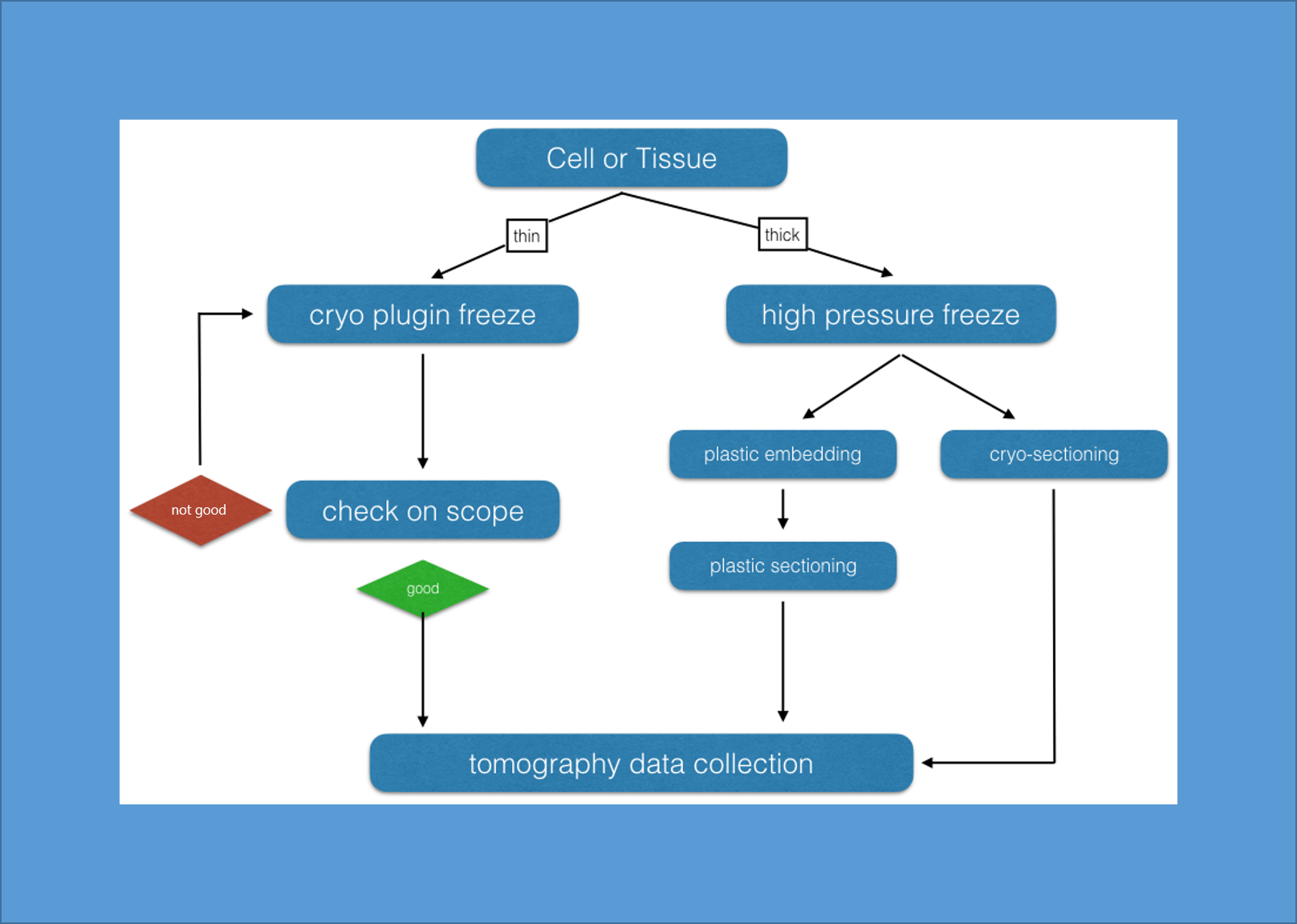
Below are two pipelines for cryo-EM specimen preparation for both Single Particle and Electron Tomography applications.
Single Particle
|
Tomography
|


