Imaging of adoptive cell transfer
Non-invasive imaging of endothelial cell (EC) surface markers holds promise in identifying early signs of inflammation, angiogenesis or atherosclerosis in biological systems.

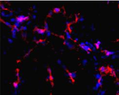
Branching of human endothelial cells formed in vivo in MatrigelTM and mouse supporting cells. CD31 staining (red fluorescence), nuclei stained with DAPI (blue).Springer-Verlag ©
We previously devised a report of E-selectin expression based on nano-sized iron oxide particles linked to a highly specific, high affinity anti-human E-selectin antibody fragment.
The experiments with targeted imaging agent suggested that the conjugates were specifically bound to EC only if marker expression was specifically induced. Specific binding of particles to EC resulted in a strong MR T2-weighted signal change. This suggested that targeted MR imaging detects inducible expression of cell-adhesion molecules.
We are currently investigating:
1. xenogeneic blood neovessels
2. targeted MR probes with the specificity against human endothelial surface markers
3. monitoring of vascularization in engineered artificial tissue grafts