Ehlers-Danlos Syndromes (EDS)
Ehlers-Danlos syndromes (EDS) are a heterogeneous group of 13 heritable connective tissue disorders first described by Hippocrates in 400 B.C. Patients with EDS have mutations in genes associated with collagen or with collagen processing, folding, or stabilization within the extracellular matrix. While signs and symptoms vary widely among the different types of EDS, most patients present with some degree of joint hypermobility, hyperextensible (“stretchy”) or fragile skin, and fragility of the blood vessels and organs. Some types of EDS are associated with life threatening complications and/or shortened life expectancy, and all are associated with a high degree of chronic pain. Currently, there is no definitive treatment for EDS.
Naturally occurring forms of EDS are found in horses, cattle, sheep, dogs, cats, rabbits, and mink. Many of these animals develop clinical signs which are quite similar to those observed in humans, making them ideal models to study the disorder.
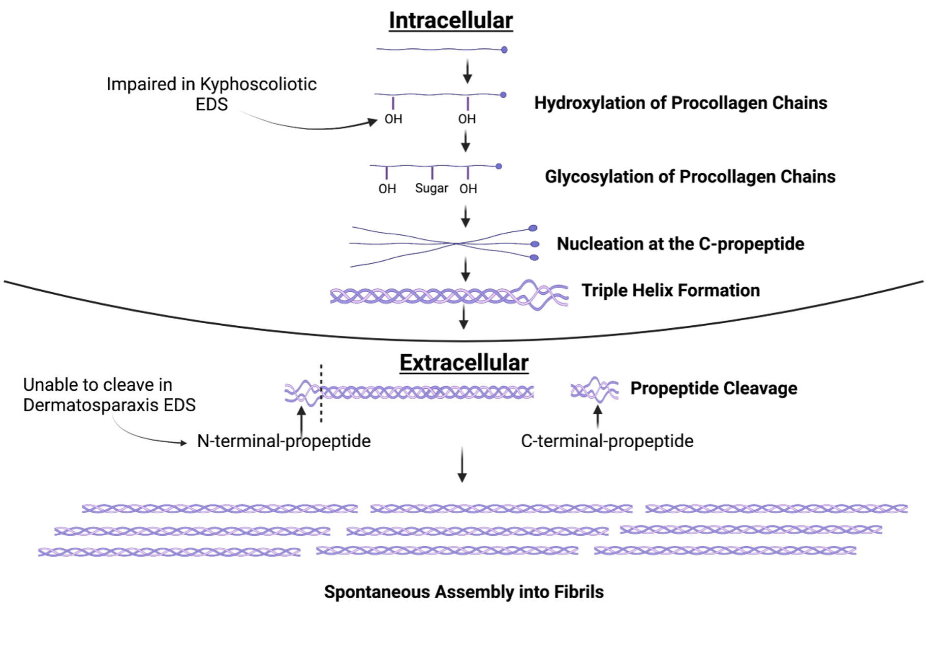
Dermatosparaxis EDS is a rare autosomal recessive disorder caused by a mutation in the gene ADAM Metallopeptidase with Thrombospondin Type 1 Motif 2 (ADAMTS2) and is found in humans, cattle, sheep, dogs, and cats. The ADAMTS2 gene encodes for the protein procollagen I N-proteinase which is responsible for cleaving the N-propeptide of procollagen chains. Mutations in this gene prevent the correct assembly of collagen fibrils. Patients with dermatosparaxis EDS present with severe skin fragility and skin laxity, easy bruising and bleeding, and preterm birth. Characteristic facial features, short stature, cerebral hemorrhages, congenital skull fractures, delayed gross motor skills such as crawling and walking, fragility of the internal organs, and umbilical hernia have also been described.
Kyphoscoliotic EDS (kEDS) is a rare autosomal recessive disorder caused by a mutation in the gene Procollagen-Lysine,2-Oxoglutarate 5-Dioxygenase 1 (PLOD1). Mutations in PLOD1 are found in both humans and horses. The PLOD1 gene encodes for the enzyme lysyl hydroxylase 1 which is responsible for hydroxylation of procollagen chains, a step needed to provide attachment points for the sugar molecules necessary for formation of the helical structure of collagen. Patients with kEDS present with severe curvature of the spine, decreased muscle tone, and joint hypermobility. They may also experience skin hyperextensibility and fragility, and aneurysms among many other complications. Currently, our lab is focused on developing CRISPR and AAV mediated gene therapies for both dermatosparaxis EDS and kyphoscoliotic EDS in mouse models.

To contact us about EDS, please email Dr. Abby McElroy at abigail.mcelroy@umassmed.edu. Please be advised, our lab does not treat or advise on the treatment of human EDS patients. Dr. McElroy is happy to discuss diagnosis and treatment of veterinary EDS patients.